Science News
Nikon Small World contest reveals unseen microscopic world all around us
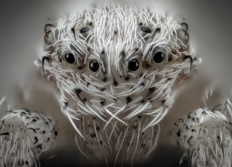
Count the eyes of a spider, investigate the inner workings of an alligator embryo and see more of the world under a microscope.
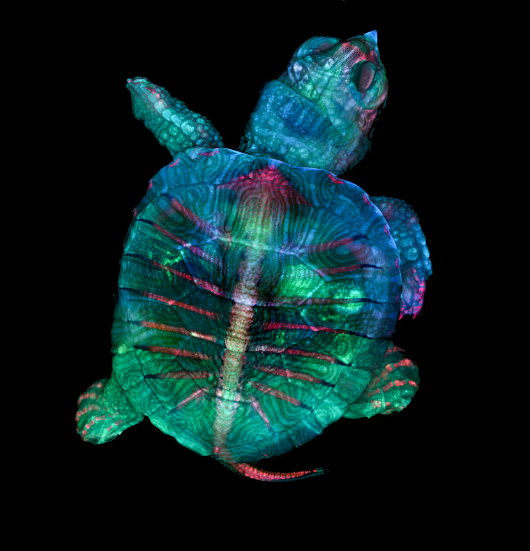
First Place
Science and art meet under the microscope in Nikon's annual Small World contest. This year a stunning fluorescent view of a turtle embryo won first place.
Microscopy technician Teresa Zgoda and recent university graduate Teresa Kugler captured the image using fluorescence and stereo microscopy.
The winning pair stacked and stitched together hundreds of images to create the final image of their turtle.
“Microscopy lets us zoom in on the smallest organisms and building blocks that comprise our world–giving us a profound appreciation for the small things in life that far too often go unnoticed,” said Kugler. “It allows me to do science with a purpose.”
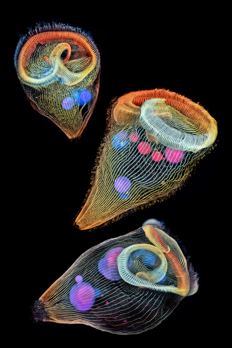
Second Place
Second place was awarded to Small World veteran Dr. Igor Siwanowicz for his composite image of three single-cell freshwater protozoans, sometimes called "trumpet animalcules.” He used confocal microscopy to capture the detail of the cilia, tiny hairs used by the animals for feeding and locomotion.
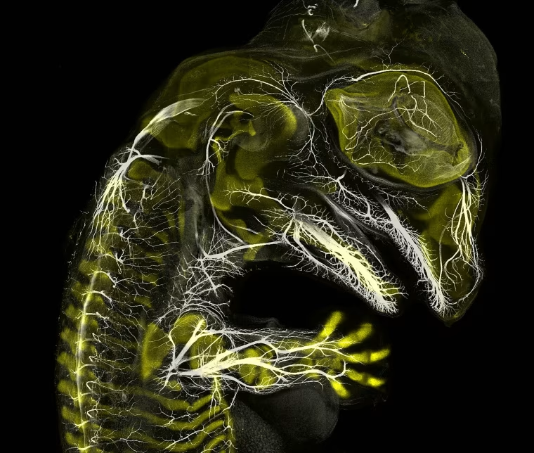
Third Place
In third place is Daniel Smith Paredes, for his image of a developing American alligator embryo. Paredes, who is studying the development and evolution of vertebrate anatomy, captured this photo at around 20 days of development using immunofluorescence.
Scroll through to see more of the top 20 winners in this year's contest.